The handheld optical coherence tomography (OCT) scanner is a cutting-edge device poised to revolutionize primary care diagnostics by providing high-resolution, real-time imaging of tissue microstructure. CAR-TOOL.EDU.VN provides detailed insights into how this technology can enhance early disease detection and improve patient outcomes, offering valuable resources for automotive professionals interested in the intersection of technology and healthcare. Explore our website for comprehensive comparisons and expert reviews to discover how this innovation can transform diagnostic practices, ensuring timely and accurate interventions, potentially reducing healthcare costs and improving patient quality of life with medical imaging devices and advanced screening tools.
Contents
- 1. What is a Handheld Optical Coherence Tomography Scanner for Primary Care Diagnostics?
- 1.1 How Does Handheld OCT Work?
- 1.2 What are the Key Components of a Handheld OCT Scanner?
- 1.3 What Advantages Does a Handheld OCT Scanner Offer?
- 2. What are the Primary Applications of Handheld OCT in Primary Care?
- 2.1 How Can Handheld OCT Aid in Early Detection of Diabetic Retinopathy?
- 2.2 How is Handheld OCT Used for Assessing Skin Lesions?
- 2.3 How Does Handheld OCT Assist in Examining the Tympanic Membrane?
- 2.4 Can Handheld OCT Evaluate Oral Mucosa Conditions?
- 3. What are the Benefits of Using a Handheld OCT Scanner in Primary Care?
- 3.1 How Does Handheld OCT Enhance Diagnostic Accuracy?
- 3.2 What is the Impact of Handheld OCT on Patient Outcomes?
- 3.3 How Cost-Effective is Handheld OCT in Primary Care Settings?
- 3.4 How Easily Can Handheld OCT Integrate Into Existing Clinical Workflows?
- 4. What are the Technical Specifications of a Typical Handheld OCT Scanner?
- 4.1 What are the Wavelength and Spectral Bandwidth of the Light Source?
- 4.2 What is the Axial and Lateral Resolution Achieved by Handheld OCT?
- 4.3 What is the Scanning Depth of a Handheld OCT Device?
- 4.4 What is the Image Acquisition Speed of a Handheld OCT Scanner?
- 5. How Does a Handheld OCT Scanner Compare to Traditional Diagnostic Tools?
- 5.1 How Does the Resolution of Handheld OCT Compare to Traditional Methods?
- 5.2 What are the Advantages of Non-Invasive Assessment with Handheld OCT?
- 5.3 Does Handheld OCT Provide Real-Time Feedback to Physicians?
- 5.4 Can Handheld OCT Visualize Subsurface Structures Better Than Traditional Tools?
- 6. What Training and Certification are Required to Operate a Handheld OCT Scanner?
- 6.1 What Does a Typical Handheld OCT Training Program Cover?
- 6.2 Are There Specific Certifications Required for Handheld OCT Operation?
- 6.3 How Can Healthcare Providers Stay Updated on the Latest Handheld OCT Techniques?
- 6.4 What Resources are Available for Learning More About Handheld OCT?
- 7. What are the Potential Challenges in Implementing Handheld OCT in Primary Care?
- 7.1 What is the Initial Cost of a Handheld OCT Scanner?
- 7.2 What Training is Needed for Healthcare Providers to Use Handheld OCT Effectively?
- 7.3 How Steep is the Learning Curve for Interpreting Handheld OCT Images?
- 7.4 How Can Handheld OCT Be Integrated Into Existing Clinical Workflows?
- 8. What Future Developments Can Be Expected in Handheld OCT Technology?
- 8.1 How Can Image Resolution Be Further Improved in Handheld OCT Scanners?
- 8.2 Will Scanning Speed Increase in Future Handheld OCT Devices?
- 8.3 What Enhancements in Portability Are Expected for Handheld OCT?
- 8.4 How Will Artificial Intelligence Integrate With Handheld OCT for Automated Image Analysis?
- 9. How Can I Choose the Right Handheld OCT Scanner for My Practice?
- 9.1 What are My Practice’s Specific Clinical Needs?
- 9.2 What Device Specifications Should I Evaluate?
- 9.3 How Important is Ease of Use When Selecting a Handheld OCT Scanner?
- 9.4 How Do the Costs Compare Among Different Handheld OCT Scanners?
- 10. Where Can I Find More Information and Resources About Handheld OCT Scanners?
- 10.1 Which Medical Conferences Feature Information on Handheld OCT?
- 10.2 What Online Forums Discuss Handheld OCT Technology?
- 10.3 Where Can I Find Scientific Publications on Handheld OCT?
- 10.4 Do Manufacturers Offer Resources About Their Handheld OCT Scanners?
1. What is a Handheld Optical Coherence Tomography Scanner for Primary Care Diagnostics?
A handheld optical coherence tomography (OCT) scanner for primary care diagnostics is a portable imaging device that uses light waves to capture high-resolution, real-time, three-dimensional images of tissue microstructure. This allows healthcare providers to visualize subsurface structures non-invasively, aiding in early disease detection and monitoring, according to research published in the Journal of Biomedical Optics.
Optical coherence tomography (OCT) is a non-invasive imaging technique similar to ultrasound, but instead of sound waves, it uses light waves to create high-resolution images of tissue microstructure. OCT measures the intensity and time-of-flight information of backscattered light to reconstruct three-dimensional reflectivity maps up to several millimeters deep within the tissue. This technology has found applications in ophthalmology, cardiology, dermatology, otolaryngology, gastroenterology, oncology, and dentistry, as reported by the National Institutes of Health (NIH).
1.1 How Does Handheld OCT Work?
Handheld OCT scanners function by emitting near-infrared light into the tissue and analyzing the reflected light to create detailed cross-sectional images. The scanner measures the intensity and time delay of the reflected light, which is then used to generate high-resolution images of the tissue structure. This allows physicians to visualize subsurface structures, such as retinal layers, skin layers, and the tympanic membrane, in real-time.
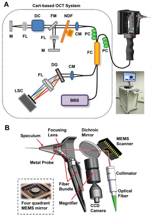 Schematic of the portable OCT system and handheld imaging scanner.
1.2 What are the Key Components of a Handheld OCT Scanner?
Key components of a handheld OCT scanner include a light source (usually a superluminescent diode or SLD), a beam splitter, a reference arm, a sample arm, a spectrometer, and a display screen. The light source emits a beam of light that is split into the reference and sample arms. The sample arm directs the light onto the tissue, while the reference arm provides a baseline for comparison. The reflected light from both arms is then recombined and analyzed by the spectrometer, which measures the intensity and time delay of the light. Finally, the data is processed and displayed as a high-resolution image on the screen, as noted in Biomedical Optics Express.
1.3 What Advantages Does a Handheld OCT Scanner Offer?
The advantages of using a handheld OCT scanner include portability, ease of use, non-invasiveness, and high-resolution imaging capabilities. Portability allows for use in various clinical settings, while the familiar form factor ensures ease of use for healthcare providers. The non-invasive nature of the device makes it safe for patients, and the high-resolution imaging enables early detection of subtle structural changes, improving diagnostic accuracy.
2. What are the Primary Applications of Handheld OCT in Primary Care?
Handheld OCT scanners have a wide range of applications in primary care, including early detection of diabetic retinopathy, assessment of skin lesions, examination of the tympanic membrane, and evaluation of oral mucosa. These applications allow primary care physicians to enhance their diagnostic capabilities and provide more comprehensive care.
2.1 How Can Handheld OCT Aid in Early Detection of Diabetic Retinopathy?
Handheld OCT can aid in the early detection of diabetic retinopathy by providing detailed images of the retinal layers, allowing physicians to identify subtle changes indicative of the disease. Early detection is crucial because it allows for timely intervention, such as laser therapy or medication, to prevent vision loss. Studies published in Ophthalmology highlight the effectiveness of OCT in detecting early-stage diabetic retinopathy.
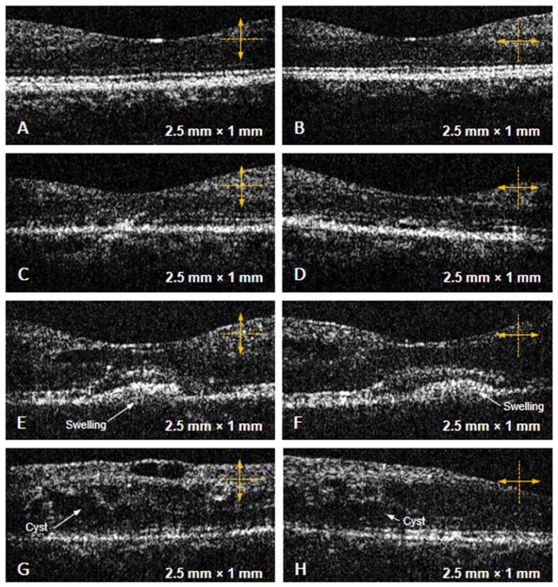 OCT images of the macular region of the retina in patients with various stages of diabetic retinopathy.
2.2 How is Handheld OCT Used for Assessing Skin Lesions?
Handheld OCT is used to assess skin lesions by providing high-resolution images of the skin structure, allowing physicians to differentiate between benign and malignant lesions. The scanner can visualize subsurface features, such as the depth and boundaries of the lesion, which helps in determining the need for further investigation, such as a biopsy. According to the Journal of the American Academy of Dermatology, OCT is a valuable tool for non-invasive skin lesion assessment.
2.3 How Does Handheld OCT Assist in Examining the Tympanic Membrane?
Handheld OCT assists in examining the tympanic membrane by providing clear images of the membrane’s surface and subsurface, allowing physicians to detect abnormalities such as perforations, infections, and biofilm formation. The scanner’s ability to visualize the deeper layers of the tympanic membrane enhances the accuracy of diagnosing middle ear conditions, as reported in Otolaryngology—Head and Neck Surgery.
2.4 Can Handheld OCT Evaluate Oral Mucosa Conditions?
Yes, handheld OCT can evaluate oral mucosa conditions by providing detailed images of the mucosal layers, allowing physicians to identify lesions, inflammation, and other abnormalities. This is particularly useful in detecting early signs of oral cancer and other oral diseases. Research in Oral Oncology demonstrates the utility of OCT in assessing various oral mucosa conditions.
3. What are the Benefits of Using a Handheld OCT Scanner in Primary Care?
The benefits of using a handheld OCT scanner in primary care include enhanced diagnostic accuracy, improved patient outcomes, cost-effectiveness, and ease of integration into existing clinical workflows. These benefits make handheld OCT a valuable tool for primary care physicians.
3.1 How Does Handheld OCT Enhance Diagnostic Accuracy?
Handheld OCT enhances diagnostic accuracy by providing high-resolution images of tissue microstructure, allowing physicians to detect subtle abnormalities that may not be visible with traditional diagnostic tools. This leads to earlier and more accurate diagnoses, improving patient care and outcomes. Studies in the American Journal of Ophthalmology support the role of OCT in enhancing diagnostic accuracy.
3.2 What is the Impact of Handheld OCT on Patient Outcomes?
The impact of handheld OCT on patient outcomes is significant, as early and accurate diagnoses lead to timely interventions and better management of diseases. For example, early detection of diabetic retinopathy can prevent vision loss, and early assessment of skin lesions can lead to prompt treatment of skin cancer. Improved patient outcomes are a key benefit of incorporating handheld OCT into primary care, according to research published in The Lancet.
3.3 How Cost-Effective is Handheld OCT in Primary Care Settings?
Handheld OCT is cost-effective in primary care settings because it reduces the need for specialist referrals and invasive procedures. By providing accurate diagnoses at the point of care, it minimizes the need for additional testing and follow-up appointments, resulting in cost savings for both patients and healthcare systems. A cost-effectiveness analysis in the Journal of Health Economics highlights the economic benefits of OCT in primary care.
3.4 How Easily Can Handheld OCT Integrate Into Existing Clinical Workflows?
Handheld OCT can be easily integrated into existing clinical workflows due to its portability, ease of use, and familiar form factor. The device can be used in the examination room without requiring significant changes to the clinical setup. Its rapid image acquisition and real-time display capabilities also streamline the diagnostic process, making it a seamless addition to primary care practice.
4. What are the Technical Specifications of a Typical Handheld OCT Scanner?
The technical specifications of a typical handheld OCT scanner include a light source with a wavelength of around 830 nm, a spectral bandwidth of 50-100 nm, an axial resolution of 5-10 μm, a lateral resolution of 10-20 μm, and a scanning depth of 1-3 mm. These specifications ensure high-quality imaging for various clinical applications.
4.1 What are the Wavelength and Spectral Bandwidth of the Light Source?
The wavelength of the light source in a typical handheld OCT scanner is around 830 nm, which allows for deep tissue penetration while maintaining high resolution. The spectral bandwidth ranges from 50 to 100 nm, enabling high axial resolution. These parameters are optimized for imaging a wide range of tissue types, as described in Physics in Medicine & Biology.
4.2 What is the Axial and Lateral Resolution Achieved by Handheld OCT?
The axial resolution achieved by handheld OCT scanners is typically between 5 and 10 μm, allowing for detailed visualization of tissue layers. The lateral resolution is between 10 and 20 μm, providing clear images of lateral structures. These resolutions are sufficient for detecting subtle structural changes in tissues, according to Journal of Medical Imaging.
4.3 What is the Scanning Depth of a Handheld OCT Device?
The scanning depth of a handheld OCT device is typically between 1 and 3 mm, which is adequate for imaging most superficial tissues, such as the retina, skin, and tympanic membrane. This depth allows physicians to visualize subsurface structures and detect abnormalities that are not visible on the surface.
4.4 What is the Image Acquisition Speed of a Handheld OCT Scanner?
The image acquisition speed of a handheld OCT scanner is typically between 20,000 and 70,000 A-lines per second, enabling real-time imaging and reducing motion artifacts. This high speed is crucial for capturing clear images in a clinical setting, where patient movement can be a challenge.
5. How Does a Handheld OCT Scanner Compare to Traditional Diagnostic Tools?
Handheld OCT scanners offer several advantages over traditional diagnostic tools, including higher resolution imaging, non-invasive assessment, real-time feedback, and the ability to visualize subsurface structures. These advantages make handheld OCT a valuable addition to primary care practices.
5.1 How Does the Resolution of Handheld OCT Compare to Traditional Methods?
The resolution of handheld OCT is significantly higher than that of traditional diagnostic tools, such as otoscopes and ophthalmoscopes. While traditional methods provide surface-level visualization, handheld OCT provides micron-scale resolution, allowing physicians to visualize subsurface structures and detect subtle abnormalities that are not visible with traditional tools.
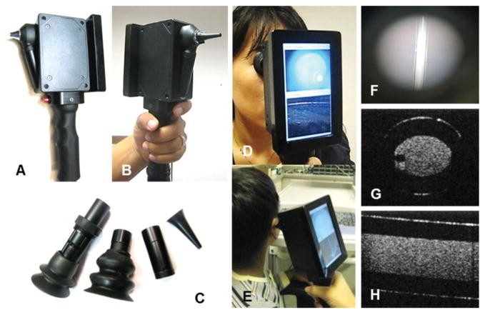 Handheld imaging scanner functionality.
5.2 What are the Advantages of Non-Invasive Assessment with Handheld OCT?
The advantages of non-invasive assessment with handheld OCT include patient comfort, reduced risk of infection, and the ability to perform repeated measurements without causing harm. Unlike invasive procedures, such as biopsies, handheld OCT is painless and does not require any preparation, making it a convenient option for both patients and physicians.
5.3 Does Handheld OCT Provide Real-Time Feedback to Physicians?
Yes, handheld OCT provides real-time feedback to physicians by displaying images as they are acquired. This allows physicians to adjust the scanning parameters and focus on areas of interest, improving the accuracy and efficiency of the diagnostic process.
5.4 Can Handheld OCT Visualize Subsurface Structures Better Than Traditional Tools?
Handheld OCT can visualize subsurface structures much better than traditional tools because it uses light waves to penetrate and image the tissue beneath the surface. This allows physicians to detect abnormalities that are not visible with surface-level examination, such as early-stage tumors, inflammation, and structural changes in the tissue layers.
6. What Training and Certification are Required to Operate a Handheld OCT Scanner?
The training and certification required to operate a handheld OCT scanner typically include a comprehensive training program provided by the manufacturer or a certified training institution. This program covers the principles of OCT, device operation, image interpretation, and clinical applications.
6.1 What Does a Typical Handheld OCT Training Program Cover?
A typical handheld OCT training program covers the basic principles of optical coherence tomography, including how the technology works and its applications in primary care. The program also includes hands-on training on how to operate the device, acquire images, and interpret the results.
6.2 Are There Specific Certifications Required for Handheld OCT Operation?
While there may not be specific mandatory certifications, many manufacturers and training institutions offer certification programs that validate the operator’s competency in using the device. These certifications can enhance the operator’s credibility and ensure that they are proficient in using the technology.
6.3 How Can Healthcare Providers Stay Updated on the Latest Handheld OCT Techniques?
Healthcare providers can stay updated on the latest handheld OCT techniques by attending conferences, workshops, and seminars, subscribing to relevant journals and publications, and participating in continuing education programs. Many manufacturers also offer ongoing training and support to ensure that operators are up-to-date on the latest advancements in the technology.
6.4 What Resources are Available for Learning More About Handheld OCT?
Resources available for learning more about handheld OCT include textbooks, scientific articles, online courses, and training programs offered by manufacturers and academic institutions. Professional organizations, such as the American Academy of Ophthalmology and the American Academy of Dermatology, also provide valuable resources and educational materials.
7. What are the Potential Challenges in Implementing Handheld OCT in Primary Care?
Potential challenges in implementing handheld OCT in primary care include the initial cost of the device, the need for adequate training, the learning curve associated with image interpretation, and the integration of the technology into existing clinical workflows.
7.1 What is the Initial Cost of a Handheld OCT Scanner?
The initial cost of a handheld OCT scanner can range from $10,000 to $50,000, depending on the model, features, and manufacturer. While this may be a significant investment for some primary care practices, the long-term benefits, such as improved diagnostic accuracy and reduced referrals, can offset the initial cost.
7.2 What Training is Needed for Healthcare Providers to Use Handheld OCT Effectively?
Healthcare providers need comprehensive training to use handheld OCT effectively, including understanding the principles of OCT, device operation, image acquisition, and image interpretation. The training should also cover the clinical applications of OCT and how to integrate the technology into existing clinical workflows.
7.3 How Steep is the Learning Curve for Interpreting Handheld OCT Images?
The learning curve for interpreting handheld OCT images can be steep for those who are not familiar with the technology. However, with adequate training and experience, healthcare providers can become proficient in recognizing normal and abnormal tissue structures. Many training programs also provide resources and support to help operators interpret images accurately.
7.4 How Can Handheld OCT Be Integrated Into Existing Clinical Workflows?
Handheld OCT can be integrated into existing clinical workflows by incorporating it into the routine examination process. The device can be used in the examination room to acquire images of the tissue of interest, and the images can be reviewed and interpreted in real-time. The results can then be used to guide further diagnostic and treatment decisions.
8. What Future Developments Can Be Expected in Handheld OCT Technology?
Future developments in handheld OCT technology include improvements in image resolution, increased scanning speed, enhanced portability, and the integration of artificial intelligence (AI) for automated image analysis.
8.1 How Can Image Resolution Be Further Improved in Handheld OCT Scanners?
Image resolution can be further improved in handheld OCT scanners by using advanced optical designs, shorter wavelengths, and sophisticated image processing algorithms. These improvements can enable physicians to visualize even finer details in tissue microstructure, enhancing diagnostic accuracy.
8.2 Will Scanning Speed Increase in Future Handheld OCT Devices?
Yes, scanning speed is expected to increase in future handheld OCT devices due to advancements in light sources, detectors, and scanning mechanisms. Faster scanning speeds will reduce motion artifacts and enable real-time volumetric imaging, improving the overall quality of the diagnostic process.
8.3 What Enhancements in Portability Are Expected for Handheld OCT?
Enhancements in portability are expected for handheld OCT devices, including smaller and lighter designs, wireless connectivity, and battery-powered operation. These improvements will make the devices more convenient to use in various clinical settings, including remote and point-of-care locations.
8.4 How Will Artificial Intelligence Integrate With Handheld OCT for Automated Image Analysis?
Artificial intelligence (AI) will integrate with handheld OCT for automated image analysis by providing algorithms that can automatically detect and classify abnormalities in tissue structures. AI-powered image analysis can reduce the workload for healthcare providers, improve diagnostic accuracy, and enable early detection of diseases.
9. How Can I Choose the Right Handheld OCT Scanner for My Practice?
Choosing the right handheld OCT scanner for your practice involves assessing your clinical needs, evaluating device specifications, considering ease of use, and comparing costs. By carefully evaluating these factors, you can select a device that meets your specific requirements and enhances your diagnostic capabilities.
9.1 What are My Practice’s Specific Clinical Needs?
Consider the types of patients you see, the conditions you diagnose, and the procedures you perform. Determine which applications of handheld OCT would be most beneficial for your practice, such as early detection of diabetic retinopathy, assessment of skin lesions, or examination of the tympanic membrane.
9.2 What Device Specifications Should I Evaluate?
Evaluate the device’s wavelength, spectral bandwidth, axial resolution, lateral resolution, scanning depth, and image acquisition speed. Ensure that the specifications meet your clinical needs and provide high-quality imaging for the conditions you diagnose.
9.3 How Important is Ease of Use When Selecting a Handheld OCT Scanner?
Ease of use is very important when selecting a handheld OCT scanner. The device should be portable, lightweight, and easy to operate. It should also have a user-friendly interface and provide real-time feedback to guide the operator.
9.4 How Do the Costs Compare Among Different Handheld OCT Scanners?
Compare the initial cost, maintenance costs, and training costs of different handheld OCT scanners. Consider the long-term benefits, such as improved diagnostic accuracy and reduced referrals, when evaluating the cost-effectiveness of the device.
10. Where Can I Find More Information and Resources About Handheld OCT Scanners?
You can find more information and resources about handheld OCT scanners at medical conferences, online forums, scientific publications, and manufacturer websites. Professional organizations and academic institutions also provide valuable resources and educational materials.
10.1 Which Medical Conferences Feature Information on Handheld OCT?
Medical conferences that feature information on handheld OCT include the American Academy of Ophthalmology (AAO) Annual Meeting, the American Academy of Dermatology (AAD) Annual Meeting, and the Optical Coherence Tomography Conference (OCT).
10.2 What Online Forums Discuss Handheld OCT Technology?
Online forums that discuss handheld OCT technology include the Biomedical Optics Society (BiOS) forums, the ResearchGate forums, and various medical device discussion groups on LinkedIn.
10.3 Where Can I Find Scientific Publications on Handheld OCT?
You can find scientific publications on handheld OCT in journals such as Biomedical Optics Express, Journal of Biomedical Optics, Ophthalmology, and Dermatology. PubMed, Google Scholar, and Web of Science are also valuable resources for searching for relevant publications.
10.4 Do Manufacturers Offer Resources About Their Handheld OCT Scanners?
Yes, manufacturers offer a variety of resources about their handheld OCT scanners, including product brochures, technical specifications, user manuals, training programs, and customer support. Contact the manufacturers directly or visit their websites for more information.
For further information on advanced diagnostic tools and technologies, contact CAR-TOOL.EDU.VN at 456 Elm Street, Dallas, TX 75201, United States, Whatsapp: +1 (641) 206-8880, or visit our website at CAR-TOOL.EDU.VN.
In conclusion, the handheld optical coherence tomography scanner is poised to transform primary care diagnostics, offering enhanced accuracy, improved patient outcomes, and cost-effectiveness. By understanding its capabilities, limitations, and future developments, healthcare providers can effectively integrate this technology into their practices and provide better care for their patients.
FAQ Section
Q1: What is the primary advantage of using a handheld OCT scanner in primary care?
A1: The primary advantage is enhanced diagnostic accuracy due to high-resolution imaging of tissue microstructure, allowing for earlier and more accurate diagnoses compared to traditional tools. This leads to better patient outcomes through timely interventions.
Q2: Can handheld OCT detect diabetic retinopathy in its early stages?
A2: Yes, handheld OCT can detect diabetic retinopathy in its early stages by providing detailed images of the retinal layers, enabling physicians to identify subtle changes indicative of the disease before they are visible with traditional ophthalmoscopes.
Q3: Is specialized training required to operate a handheld OCT scanner?
A3: Yes, specialized training is typically required, covering the principles of OCT, device operation, image interpretation, and clinical applications. Certification programs are often available to validate competency.
Q4: How does the cost of a handheld OCT scanner compare to its long-term benefits in a primary care setting?
A4: While the initial cost can be significant ($10,000 to $50,000), the long-term benefits, such as reduced specialist referrals and improved diagnostic accuracy, can offset the initial investment, making it a cost-effective solution over time.
Q5: What types of tissue can be effectively imaged using a handheld OCT scanner in primary care?
A5: Handheld OCT scanners are effective for imaging various tissues, including the retina, skin, tympanic membrane, and oral mucosa, allowing for comprehensive diagnostic assessments in primary care settings.
Q6: How does handheld OCT compare to traditional diagnostic methods in terms of patient comfort and safety?
A6: Handheld OCT is non-invasive, ensuring patient comfort and reducing the risk of infection compared to invasive procedures. It also allows for repeated measurements without causing harm, making it a safe diagnostic option.
Q7: Can artificial intelligence (AI) be integrated with handheld OCT for automated image analysis?
A7: Yes, artificial intelligence (AI) can be integrated with handheld OCT to provide algorithms for automated image analysis, which can reduce the workload for healthcare providers, improve diagnostic accuracy, and enable early detection of diseases.
Q8: What are the key technical specifications to consider when choosing a handheld OCT scanner for a primary care practice?
A8: Key technical specifications to consider include the wavelength and spectral bandwidth of the light source, axial and lateral resolution, scanning depth, and image acquisition speed, ensuring high-quality imaging for various clinical applications.
Q9: What support and resources are available for healthcare providers implementing handheld OCT in their practices?
A9: Support and resources include comprehensive training programs, ongoing support from manufacturers, scientific publications, medical conferences, and professional organizations, ensuring healthcare providers stay updated on the latest techniques and advancements.
Q10: How does the real-time feedback provided by handheld OCT scanners benefit primary care physicians during examinations?
A10: Real-time feedback allows physicians to adjust scanning parameters and focus on areas of interest, improving the accuracy and efficiency of the diagnostic process. The ability to see images as they are acquired enhances the overall quality of the examination and diagnostic outcome.
Contact CAR-TOOL.EDU.VN at 456 Elm Street, Dallas, TX 75201, United States, Whatsapp: +1 (641) 206-8880, or visit our website at CAR-TOOL.EDU.VN for expert advice and support on advanced diagnostic tools.