Southeast Breast Care & Diagnostic Doctors Park in Cape Girardeau, MO, provides a range of services focused on breast health and diagnostics. CAR-TOOL.EDU.VN helps you understand what this entails, including the types of care offered, the technologies used, and why it’s a crucial resource for the community. Find out more about advanced breast imaging and diagnostic services, ensuring you or your loved ones receive the best possible care by discussing treatment options.
Contents
- 1. What Is Southeast Breast Care & Diagnostic Doctors Park Cape Girardeau MO?
- 1.1 Comprehensive Breast Health Services
- 1.2 Advanced Imaging Technologies
- 1.3 Personalized Care Approach
- 1.4 Multidisciplinary Team
- 1.5 Importance of Early Detection
- 1.6 Location and Accessibility
- 1.7 Commitment to Excellence
- 1.8 Community Resource
- 1.9 CAR-TOOL.EDU.VN Partnership
- 1.10 Further Information
- 2. What Are The Key Services Offered?
- 2.1 Screening Mammography
- 2.2 Diagnostic Mammography
- 2.3 Breast Ultrasound
- 2.4 Breast MRI (Magnetic Resonance Imaging)
- 2.5 Breast Biopsy
- 2.6 Image-Guided Biopsy
- 2.7 Risk Assessment and Genetic Counseling
- 2.8 Second Opinion Services
- 2.9 Follow-Up Care and Monitoring
- 2.10 Support Services
- 3. How Does Advanced Imaging Aid in Diagnosis?
- 3.1 3D Mammography (Tomosynthesis)
- 3.2 High-Resolution Ultrasound
- 3.3 Breast MRI (Magnetic Resonance Imaging)
- 3.4 Contrast-Enhanced Mammography (CEM)
- 3.5 Molecular Breast Imaging (MBI)
- 3.6 Automated Breast Ultrasound (ABUS)
- 3.7 Artificial Intelligence (AI) in Breast Imaging
- 3.8 Elastography
- 3.9 Thermography
- 3.10 Positron Emission Tomography (PET) Scans
- 4. What Should I Expect During My First Visit?
- 4.1 Registration and Paperwork
- 4.2 Medical History Review
- 4.3 Physical Examination
- 4.4 Discussion of Symptoms and Concerns
- 4.5 Diagnostic Imaging
- 4.6 Mammography
- 4.7 Ultrasound
- 4.8 MRI
- 4.9 Review of Findings and Recommendations
- 4.10 Scheduling Follow-Up Appointments
- 5. How Can I Prepare For My Appointment?
- 5.1 Gather Relevant Medical Records
- 5.2 List of Medications and Allergies
- 5.3 Avoid Using Deodorants, Lotions, and Powders
- 5.4 Wear Comfortable Clothing
- 5.5 Arrive on Time
- 5.6 Prepare a List of Questions
- 5.7 Inform the Clinic About Special Needs
- 5.8 Bring a Support Person
- 5.9 Review Insurance Coverage
- 5.10 Stay Hydrated
- 6. What Are the Risk Factors for Breast Cancer?
- 6.1 Age
- 6.2 Family History
- 6.3 Genetic Mutations
- 6.4 Personal History of Breast Conditions
- 6.5 Dense Breast Tissue
- 6.6 Radiation Exposure
- 6.7 Obesity
- 6.8 Alcohol Consumption
- 6.9 Hormone Therapy
- 6.10 Reproductive History
- 7. What Is The Role Of Biopsies in Diagnosis?
- 7.1 Confirmation of Cancer Diagnosis
- 7.2 Differentiation Between Benign and Malignant Tumors
- 7.3 Determination of Cancer Type and Grade
- 7.4 Evaluation of Hormone Receptor Status
- 7.5 Assessment of HER2 Status
- 7.6 Guidance for Treatment Planning
- 7.7 Monitoring Treatment Response
- 7.8 Types of Breast Biopsies
- 7.9 Image-Guided Biopsies
- 7.10 Patient Comfort and Safety
- 8. How Does Southeast Breast Care Ensure Patient Comfort and Privacy?
- 8.1 Caring and Compassionate Staff
- 8.2 Private Examination Rooms
- 8.3 Clear Communication
- 8.4 Respect for Patient Preferences
- 8.5 Adherence to HIPAA Regulations
- 8.6 Confidentiality Protocols
- 8.7 Comfortable Waiting Areas
- 8.8 Minimally Invasive Procedures
- 8.9 Pain Management Strategies
- 8.10 Support Services
- 9. What Follow-Up Care Is Typically Recommended?
- 9.1 Regular Screening Mammograms
- 9.2 Clinical Breast Exams
- 9.3 Breast Self-Exams
- 9.4 Follow-Up Imaging
- 9.5 Biopsy
- 9.6 Genetic Counseling and Testing
- 9.7 Risk-Reducing Medications
- 9.8 Lifestyle Modifications
- 9.9 Surveillance for High-Risk Individuals
- 9.10 Individualized Care Plans
- 10. How Can I Contact Southeast Breast Care & Diagnostic Doctors Park?
- 10.1 Phone Contact
- 10.2 Website Inquiry
- 10.3 Online Patient Portal
- 10.4 In-Person Visit
- 10.5 Mailing Address
- 10.6 Emergency Contact
- 10.7 Insurance Questions
- 10.8 Feedback and Complaints
- 10.9 Social Media
- 10.10 CAR-TOOL.EDU.VN Resources
- FAQ About Southeast Breast Care & Diagnostic Doctors Park Cape Girardeau MO
- 1. What types of breast imaging services are offered?
- 2. At what age should women start getting mammograms?
- 3. What is 3D mammography (tomosynthesis)?
- 4. What is breast ultrasound used for?
- 5. What is breast MRI used for?
- 6. What is a breast biopsy?
- 7. How can I prepare for my first appointment?
- 8. What are the main risk factors for breast cancer?
- 9. What is the role of biopsies in diagnosis?
- 10. How can I contact Southeast Breast Care & Diagnostic Doctors Park?
1. What Is Southeast Breast Care & Diagnostic Doctors Park Cape Girardeau MO?
Southeast Breast Care & Diagnostic Doctors Park in Cape Girardeau, MO, is a specialized healthcare facility focused on comprehensive breast health services, including advanced imaging, diagnostics, and personalized care. This clinic is dedicated to early detection, accurate diagnosis, and effective management of breast conditions.
1.1 Comprehensive Breast Health Services
The facility provides a wide array of services, including screening mammography, diagnostic mammography, breast ultrasound, breast MRI, and biopsies. These services are designed to cater to different needs, from routine check-ups to more complex diagnostic evaluations.
1.2 Advanced Imaging Technologies
Utilizing state-of-the-art equipment, the clinic ensures high-quality imaging for accurate and early detection of breast abnormalities. Technologies like 3D mammography (tomosynthesis) and high-resolution ultrasound enhance the precision of screenings and diagnostics.
1.3 Personalized Care Approach
Southeast Breast Care & Diagnostic Doctors Park emphasizes a patient-centered approach, offering individualized care plans tailored to each patient’s specific needs and medical history. This includes thorough consultations, detailed explanations of procedures, and compassionate support throughout the entire process.
1.4 Multidisciplinary Team
The clinic boasts a team of experienced radiologists, surgeons, oncologists, and support staff who collaborate to provide comprehensive care. This multidisciplinary approach ensures that patients receive well-rounded and coordinated treatment.
1.5 Importance of Early Detection
Early detection is crucial in improving outcomes for breast conditions. The clinic’s focus on advanced diagnostics and regular screenings helps identify potential issues at an early stage, allowing for more effective treatment options and improved prognosis.
1.6 Location and Accessibility
Conveniently located in Doctors Park, Cape Girardeau, the facility is easily accessible for residents of Southeast Missouri and surrounding areas. Its location ensures that patients can access high-quality breast care services without having to travel long distances.
1.7 Commitment to Excellence
The clinic is committed to maintaining the highest standards of care, continually updating its technologies and protocols to align with the latest advancements in breast health. This commitment ensures that patients receive the best possible care and outcomes.
1.8 Community Resource
As a vital healthcare resource, Southeast Breast Care & Diagnostic Doctors Park plays a significant role in promoting breast health awareness and providing accessible, high-quality services to the community.
1.9 CAR-TOOL.EDU.VN Partnership
CAR-TOOL.EDU.VN supports and promotes the clinic by providing information and resources to help individuals understand the importance of regular breast screenings and the comprehensive services available at Southeast Breast Care & Diagnostic Doctors Park.
1.10 Further Information
For more detailed information about the services offered, appointment scheduling, and insurance coverage, individuals are encouraged to visit the clinic’s website or contact them directly.
2. What Are The Key Services Offered?
Southeast Breast Care & Diagnostic Doctors Park in Cape Girardeau, MO, offers a comprehensive suite of services, including mammography, ultrasound, MRI, and biopsy procedures, to ensure early detection and accurate diagnosis of breast conditions.
2.1 Screening Mammography
Screening mammography is a key preventive service designed to detect breast cancer in its earliest stages, often before any symptoms appear. It is recommended annually for women starting at age 40, according to the American Cancer Society. Early detection through screening mammography significantly improves treatment outcomes and survival rates.
2.2 Diagnostic Mammography
Diagnostic mammography is used to investigate suspicious findings from a screening mammogram or to evaluate specific breast symptoms, such as a lump, pain, or nipple discharge. This type of mammogram provides more detailed images and may include additional views to better assess the area of concern. The process helps in determining the nature of the abnormality and guides further diagnostic steps if needed.
2.3 Breast Ultrasound
Breast ultrasound uses sound waves to create images of the breast tissue. It is often used as a supplemental imaging tool to mammography, particularly in women with dense breast tissue, where mammograms can be less effective. Ultrasound can help distinguish between solid masses and fluid-filled cysts and can guide biopsies of suspicious areas.
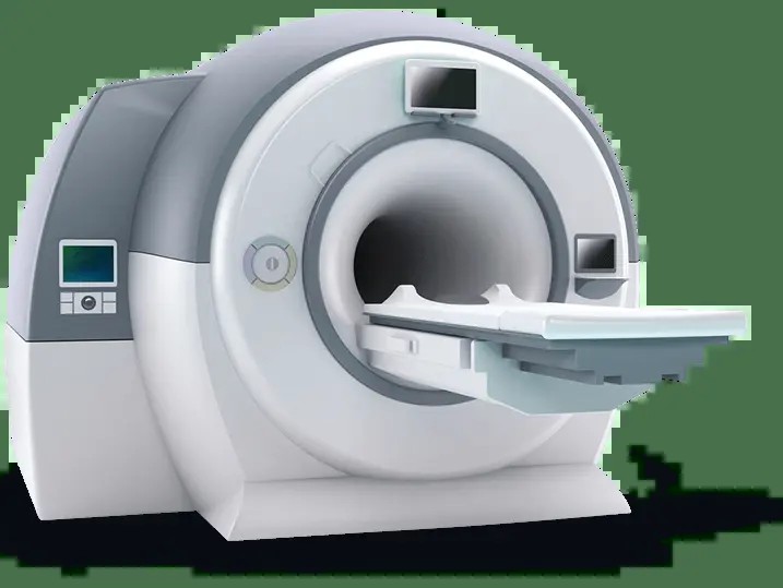
2.4 Breast MRI (Magnetic Resonance Imaging)
Breast MRI is a highly sensitive imaging technique that uses magnetic fields and radio waves to produce detailed images of the breast. It is often recommended for women at high risk of breast cancer, those with dense breast tissue, or to evaluate the extent of cancer after a diagnosis. Breast MRI can detect abnormalities that may not be visible on mammograms or ultrasounds.
2.5 Breast Biopsy
A breast biopsy involves removing a small sample of breast tissue for examination under a microscope. This procedure is performed to determine whether an abnormality is benign (non-cancerous) or malignant (cancerous). Different types of biopsies, such as needle biopsy, core biopsy, and surgical biopsy, may be used depending on the size and location of the suspicious area.
2.6 Image-Guided Biopsy
Image-guided biopsies use imaging techniques like ultrasound, mammography, or MRI to precisely locate and sample suspicious areas in the breast. This ensures that the biopsy targets the specific area of concern, improving the accuracy of the diagnosis. Image-guided biopsies are less invasive than surgical biopsies and can often be performed on an outpatient basis.
2.7 Risk Assessment and Genetic Counseling
The clinic offers risk assessment services to evaluate a woman’s risk of developing breast cancer based on factors such as family history, genetic mutations, and lifestyle. Genetic counseling is available to discuss genetic testing options and help women understand their risk and make informed decisions about screening and prevention.
2.8 Second Opinion Services
For patients seeking additional confirmation or alternative perspectives on their diagnosis or treatment plan, the clinic provides second opinion services. Expert radiologists and oncologists review the patient’s medical history, imaging studies, and biopsy results to offer a comprehensive second opinion and recommendations.
2.9 Follow-Up Care and Monitoring
The clinic provides ongoing follow-up care and monitoring for patients who have been diagnosed with breast conditions or are at high risk of developing breast cancer. This includes regular screenings, physical exams, and consultations to ensure early detection of any new or recurrent problems.
2.10 Support Services
Southeast Breast Care & Diagnostic Doctors Park offers a range of support services to help patients cope with the emotional and practical challenges of breast cancer diagnosis and treatment. These services may include counseling, support groups, educational resources, and referrals to other healthcare professionals and community organizations.
3. How Does Advanced Imaging Aid in Diagnosis?
Advanced imaging at Southeast Breast Care & Diagnostic Doctors Park in Cape Girardeau, MO, enhances diagnostic accuracy through technologies like 3D mammography, high-resolution ultrasound, and breast MRI, enabling earlier and more precise detection of breast abnormalities.
3.1 3D Mammography (Tomosynthesis)
3D mammography, also known as tomosynthesis, is an advanced form of mammography that takes multiple X-ray images of the breast from different angles. These images are then reconstructed into a three-dimensional view of the breast tissue. According to a study published in the Journal of the American Medical Association (JAMA), tomosynthesis has been shown to increase cancer detection rates and reduce false-positive results compared to traditional 2D mammography.
3.2 High-Resolution Ultrasound
High-resolution ultrasound uses sound waves to create detailed images of the breast tissue. It is particularly useful for evaluating dense breast tissue and distinguishing between solid masses and fluid-filled cysts. High-resolution ultrasound can also guide biopsies of suspicious areas, ensuring accurate sampling. The American College of Radiology recommends ultrasound as a supplemental screening tool for women with dense breasts.
3.3 Breast MRI (Magnetic Resonance Imaging)
Breast MRI is a highly sensitive imaging technique that uses magnetic fields and radio waves to produce detailed images of the breast. It is often used for women at high risk of breast cancer, those with dense breast tissue, or to evaluate the extent of cancer after a diagnosis. Breast MRI can detect abnormalities that may not be visible on mammograms or ultrasounds. A study in the New England Journal of Medicine found that breast MRI significantly improves cancer detection in high-risk women.
3.4 Contrast-Enhanced Mammography (CEM)
Contrast-enhanced mammography (CEM) involves injecting a contrast agent into the bloodstream to highlight areas of increased blood flow, which can indicate cancerous tissue. CEM is particularly useful for detecting small, aggressive tumors that may be missed by traditional mammography. Research published in Radiology suggests that CEM has a similar sensitivity to breast MRI but is more accessible and less expensive.
3.5 Molecular Breast Imaging (MBI)
Molecular breast imaging (MBI) uses a radioactive tracer to detect metabolically active cancer cells in the breast. MBI is more sensitive than mammography for detecting small tumors in dense breast tissue. A study in the American Journal of Roentgenology found that MBI detected three times as many cancers as mammography in women with dense breasts.
3.6 Automated Breast Ultrasound (ABUS)
Automated breast ultrasound (ABUS) is a screening tool specifically designed for women with dense breast tissue. ABUS uses a wide-field transducer to acquire comprehensive images of the entire breast, which are then reviewed by a radiologist. ABUS can detect additional cancers that are not visible on mammography in women with dense breasts. The FDA has approved ABUS as a supplemental screening tool for this population.
3.7 Artificial Intelligence (AI) in Breast Imaging
Artificial intelligence (AI) is increasingly being used to enhance the accuracy and efficiency of breast imaging. AI algorithms can analyze mammograms, ultrasounds, and MRIs to identify suspicious areas and assist radiologists in making diagnoses. AI has the potential to reduce false-positive and false-negative results, improve cancer detection rates, and streamline the workflow of breast imaging departments. Research published in Nature demonstrates the potential of AI to improve the accuracy of breast cancer screening.
3.8 Elastography
Elastography is an imaging technique that measures the stiffness of breast tissue. Cancerous tumors tend to be stiffer than benign tissue, so elastography can help differentiate between benign and malignant lesions. Elastography can be performed using ultrasound or MRI and can improve the specificity of breast imaging.
3.9 Thermography
Thermography uses infrared cameras to detect heat patterns on the surface of the breast. Cancerous tumors often have increased blood flow and metabolic activity, which can cause them to appear hotter than surrounding tissue. While thermography is not a substitute for mammography, it can be used as a supplemental screening tool, particularly for women who cannot undergo mammography due to radiation exposure or other concerns.
3.10 Positron Emission Tomography (PET) Scans
Positron Emission Tomography (PET) scans are advanced imaging tests that detect metabolic activity in the body’s cells, often used to identify cancer. According to Mayo Clinic, PET scans can detect diseases before other imaging techniques because they highlight changes at the cellular level.
4. What Should I Expect During My First Visit?
During your first visit to Southeast Breast Care & Diagnostic Doctors Park in Cape Girardeau, MO, expect a comprehensive assessment including a review of your medical history, a physical exam, and possibly diagnostic imaging, all conducted with a focus on comfort and clear communication.
4.1 Registration and Paperwork
Upon arrival, you will be asked to complete registration forms providing your personal information, medical history, insurance details, and consent for treatment. Ensure you bring your identification, insurance card, and any referral forms from your primary care physician. Accurate and complete paperwork helps streamline the process and ensures that the clinic has all the necessary information for your care.
4.2 Medical History Review
A healthcare professional will review your medical history, including any prior breast conditions, surgeries, family history of breast cancer, and current medications. Be prepared to answer detailed questions about your health and lifestyle, as this information is crucial for assessing your risk and determining the appropriate screening or diagnostic plan.
4.3 Physical Examination
A physical examination of your breasts will be performed to check for any lumps, skin changes, nipple discharge, or other abnormalities. The healthcare provider will also examine the lymph nodes in your underarm area. This examination provides valuable information that complements imaging studies.
4.4 Discussion of Symptoms and Concerns
You will have the opportunity to discuss any symptoms or concerns you may have about your breast health. This is an important part of the visit, as your input helps guide the diagnostic process and ensures that all your questions are addressed. Be open and honest about your symptoms, as this will help the healthcare provider provide the best possible care.
4.5 Diagnostic Imaging
Depending on your medical history, symptoms, and the results of the physical examination, you may undergo diagnostic imaging, such as mammography, ultrasound, or MRI. The specific imaging tests recommended will depend on your individual needs and risk factors. The imaging process will be explained to you in detail, and you will have the opportunity to ask questions.
4.6 Mammography
If a mammogram is recommended, a trained technologist will position your breast in the mammography machine and take X-ray images. You may experience some pressure during the procedure, but it is generally brief. The technologist will ensure that you are as comfortable as possible throughout the process.
4.7 Ultrasound
If an ultrasound is recommended, a gel will be applied to your breast, and a handheld transducer will be used to create images of the breast tissue. Ultrasound is painless and does not involve radiation. The healthcare provider will explain the findings of the ultrasound and answer any questions you may have.
4.8 MRI
If a breast MRI is recommended, you will lie face down on a table inside the MRI machine. A contrast agent may be injected into your arm to enhance the images. MRI is painless but can be time-consuming. The healthcare provider will explain the procedure and answer any questions you may have.
4.9 Review of Findings and Recommendations
After the imaging tests are completed, a radiologist will review the images and provide a report to your healthcare provider. You will then discuss the findings and recommendations, which may include further imaging, a biopsy, or a follow-up appointment. Make sure you understand the recommendations and have all your questions answered before leaving the clinic.
4.10 Scheduling Follow-Up Appointments
If additional tests or follow-up appointments are needed, the scheduling staff will assist you in making the necessary arrangements. Be sure to keep track of your appointments and follow the recommended schedule to ensure timely and appropriate care.
5. How Can I Prepare For My Appointment?
To prepare for your appointment at Southeast Breast Care & Diagnostic Doctors Park in Cape Girardeau, MO, gather relevant medical records, avoid using deodorants or lotions, and be ready to discuss your medical history and any specific concerns with your healthcare provider.
5.1 Gather Relevant Medical Records
Collect all relevant medical records, including previous mammogram reports, ultrasound reports, MRI reports, and any biopsy results. Having these records readily available will help the healthcare provider understand your medical history and make informed decisions about your care.
5.2 List of Medications and Allergies
Prepare a list of all medications you are currently taking, including prescription drugs, over-the-counter medications, and supplements. Also, note any allergies you have to medications, contrast agents, or other substances. This information is important for ensuring your safety during any procedures or imaging tests.
5.3 Avoid Using Deodorants, Lotions, and Powders
Do not use deodorants, antiperspirants, lotions, creams, or powders on your breasts or underarms on the day of your mammogram. These products can interfere with the mammogram images and may lead to false-positive results.
5.4 Wear Comfortable Clothing
Wear comfortable clothing to your appointment. You may be asked to change into a gown for certain procedures, so choose clothing that is easy to remove and put back on.
5.5 Arrive on Time
Arrive at least 15 minutes before your scheduled appointment time to allow time for registration and paperwork. Being on time ensures that you have enough time to complete all the necessary steps before your appointment.
5.6 Prepare a List of Questions
Write down any questions or concerns you have about your breast health or the procedures you will undergo. This will help you remember to ask all your questions during your appointment and ensure that you receive the information you need.
5.7 Inform the Clinic About Special Needs
If you have any special needs, such as mobility issues, visual or hearing impairments, or anxiety about medical procedures, inform the clinic when you schedule your appointment. This will allow the staff to make the necessary accommodations to ensure your comfort and safety.
5.8 Bring a Support Person
Consider bringing a support person with you to your appointment. Having a friend or family member with you can provide emotional support and help you remember important information.
5.9 Review Insurance Coverage
Check with your insurance company to understand your coverage for breast screening and diagnostic services. Knowing your coverage will help you avoid unexpected costs and make informed decisions about your care.
5.10 Stay Hydrated
Drink plenty of water in the days leading up to your appointment. Staying hydrated can help improve the quality of imaging tests and make it easier to draw blood if necessary.
6. What Are the Risk Factors for Breast Cancer?
Risk factors for breast cancer include age, family history, genetic mutations, personal history of breast conditions, and lifestyle factors such as obesity and alcohol consumption, all of which can influence the likelihood of developing the disease.
6.1 Age
The risk of breast cancer increases with age. Most breast cancers are diagnosed after age 50. According to the American Cancer Society, about two out of three invasive breast cancers are found in women 55 or older.
6.2 Family History
Having a family history of breast cancer, particularly in a mother, sister, or daughter, increases your risk. The risk is higher if your relative was diagnosed at a younger age. A study published in the Journal of the National Cancer Institute found that women with a first-degree relative (mother, sister, or daughter) with breast cancer have about a two-fold increased risk of developing the disease.
6.3 Genetic Mutations
Certain genetic mutations, such as BRCA1 and BRCA2, significantly increase the risk of breast cancer. These genes are involved in DNA repair, and mutations in these genes can lead to uncontrolled cell growth. Genetic testing can identify these mutations, allowing for more intensive screening and preventive measures.
6.4 Personal History of Breast Conditions
Having a personal history of certain benign breast conditions, such as atypical hyperplasia or lobular carcinoma in situ (LCIS), increases the risk of developing breast cancer. These conditions are not cancer but can increase the likelihood of developing cancer in the future.
6.5 Dense Breast Tissue
Women with dense breast tissue have a higher risk of breast cancer. Dense breast tissue has more glandular and fibrous tissue and less fatty tissue, making it more difficult to detect tumors on mammograms. Some states have laws requiring healthcare providers to inform women if they have dense breast tissue and discuss additional screening options.
6.6 Radiation Exposure
Exposure to radiation, particularly during childhood or adolescence, increases the risk of breast cancer. This includes radiation therapy for other cancers and exposure to atomic bomb blasts. The risk is higher with higher doses of radiation.
6.7 Obesity
Being overweight or obese, particularly after menopause, increases the risk of breast cancer. Fat tissue produces estrogen, which can promote the growth of breast cancer cells. Maintaining a healthy weight can help reduce the risk.
6.8 Alcohol Consumption
Drinking alcohol increases the risk of breast cancer. The risk increases with the amount of alcohol consumed. The American Cancer Society recommends that women limit their alcohol intake to no more than one drink per day.
6.9 Hormone Therapy
Long-term use of hormone therapy, particularly estrogen plus progestin, increases the risk of breast cancer. The risk decreases when hormone therapy is stopped. Women should discuss the risks and benefits of hormone therapy with their healthcare provider.
6.10 Reproductive History
Certain factors related to reproductive history can affect the risk of breast cancer. These include starting menstruation at an early age, having your first child at an older age, and not having children. These factors are thought to increase the lifetime exposure to estrogen, which can increase the risk of breast cancer.
7. What Is The Role Of Biopsies in Diagnosis?
Biopsies play a crucial role in breast cancer diagnosis by providing a tissue sample for microscopic examination, allowing pathologists to determine whether an abnormality is benign or malignant and guide treatment decisions.
7.1 Confirmation of Cancer Diagnosis
A biopsy is the only way to definitively confirm whether a breast abnormality is cancerous. Imaging tests like mammograms, ultrasounds, and MRIs can detect suspicious areas, but a biopsy is needed to determine the nature of the cells. The American Cancer Society emphasizes that a biopsy is essential for an accurate diagnosis.
7.2 Differentiation Between Benign and Malignant Tumors
Biopsies allow pathologists to examine tissue samples under a microscope and determine whether the cells are benign (non-cancerous) or malignant (cancerous). This differentiation is crucial for guiding treatment decisions.
7.3 Determination of Cancer Type and Grade
If cancer is present, a biopsy can determine the type of breast cancer (e.g., ductal carcinoma, lobular carcinoma) and its grade (a measure of how aggressive the cancer cells are). This information helps healthcare providers tailor treatment plans to the specific characteristics of the cancer.
7.4 Evaluation of Hormone Receptor Status
Biopsies are used to evaluate the hormone receptor status of breast cancer cells. This includes determining whether the cells have estrogen receptors (ER) and progesterone receptors (PR). Hormone receptor-positive cancers can be treated with hormone therapy, which blocks the effects of estrogen and progesterone.
7.5 Assessment of HER2 Status
Biopsies are also used to assess the HER2 (human epidermal growth factor receptor 2) status of breast cancer cells. HER2 is a protein that promotes cell growth. HER2-positive cancers can be treated with targeted therapies that block the HER2 protein.
7.6 Guidance for Treatment Planning
The information obtained from a biopsy is essential for developing an effective treatment plan. The type of cancer, its grade, hormone receptor status, and HER2 status all influence the choice of treatment options, which may include surgery, chemotherapy, radiation therapy, hormone therapy, and targeted therapy.
7.7 Monitoring Treatment Response
In some cases, biopsies may be performed during or after treatment to monitor the response of the cancer cells to therapy. This can help healthcare providers determine whether the treatment is effective and make adjustments if needed.
7.8 Types of Breast Biopsies
Several types of breast biopsies are used to obtain tissue samples for examination. These include:
- Fine-Needle Aspiration (FNA): A thin needle is used to extract fluid or cells from a suspicious area.
- Core Needle Biopsy: A larger needle is used to remove a small core of tissue.
- Incisional Biopsy: A small incision is made to remove a sample of tissue.
- Excisional Biopsy: The entire abnormal area is removed, along with some surrounding tissue.
7.9 Image-Guided Biopsies
Image-guided biopsies use imaging techniques like ultrasound, mammography, or MRI to precisely locate and sample suspicious areas in the breast. This ensures that the biopsy targets the specific area of concern, improving the accuracy of the diagnosis.
7.10 Patient Comfort and Safety
Breast biopsies are typically performed on an outpatient basis and are generally well-tolerated. Local anesthesia is used to numb the area and minimize discomfort. Patients may experience some bruising or soreness after the procedure, but this usually resolves within a few days.
8. How Does Southeast Breast Care Ensure Patient Comfort and Privacy?
Southeast Breast Care & Diagnostic Doctors Park ensures patient comfort and privacy through a caring staff, private examination rooms, clear communication, and adherence to strict confidentiality protocols.
8.1 Caring and Compassionate Staff
The clinic employs a team of healthcare professionals who are dedicated to providing compassionate and supportive care. Staff members are trained to be sensitive to the emotional and physical needs of patients and to create a welcoming and reassuring environment.
8.2 Private Examination Rooms
The clinic provides private examination rooms where patients can discuss their concerns and undergo physical examinations in a comfortable and confidential setting. These rooms are designed to ensure patient privacy and dignity.
8.3 Clear Communication
Healthcare providers at Southeast Breast Care & Diagnostic Doctors Park prioritize clear and open communication with patients. They explain procedures and treatment options in a way that is easy to understand and encourage patients to ask questions and express their concerns.
8.4 Respect for Patient Preferences
The clinic respects patient preferences and involves them in decision-making about their care. Patients are given the opportunity to express their wishes and values, and these are taken into account when developing treatment plans.
8.5 Adherence to HIPAA Regulations
The clinic adheres to the Health Insurance Portability and Accountability Act (HIPAA) regulations, which protect the privacy and security of patient health information. All patient records are kept confidential and are only shared with authorized individuals.
8.6 Confidentiality Protocols
The clinic has strict confidentiality protocols in place to ensure that patient information is protected at all times. Staff members are trained to handle patient information with the utmost care and discretion.
8.7 Comfortable Waiting Areas
The clinic provides comfortable waiting areas where patients can relax before and after their appointments. These areas are designed to be calming and welcoming, with amenities such as comfortable seating, reading materials, and refreshments.
8.8 Minimally Invasive Procedures
The clinic offers minimally invasive procedures whenever possible to reduce discomfort and speed recovery. These procedures use smaller incisions and advanced techniques to minimize trauma to the body.
8.9 Pain Management Strategies
The clinic employs a range of pain management strategies to ensure that patients are as comfortable as possible during procedures. These strategies may include local anesthesia, pain medication, and relaxation techniques.
8.10 Support Services
Southeast Breast Care & Diagnostic Doctors Park offers a range of support services to help patients cope with the emotional and practical challenges of breast cancer diagnosis and treatment. These services may include counseling, support groups, educational resources, and referrals to other healthcare professionals and community organizations.
9. What Follow-Up Care Is Typically Recommended?
Follow-up care at Southeast Breast Care & Diagnostic Doctors Park typically includes regular screenings, physical exams, and consultations, tailored to individual risk factors and previous findings, to monitor breast health and detect any changes early.
9.1 Regular Screening Mammograms
Regular screening mammograms are recommended for most women, typically starting at age 40. The frequency of mammograms may vary depending on individual risk factors and previous findings. The American Cancer Society recommends annual mammograms for women ages 45 to 54 and then every other year for women 55 and older.
9.2 Clinical Breast Exams
Clinical breast exams are performed by a healthcare provider to check for any lumps, skin changes, nipple discharge, or other abnormalities. These exams are typically performed as part of a routine check-up and can help detect potential problems early.
9.3 Breast Self-Exams
Women are encouraged to perform regular breast self-exams to become familiar with the normal look and feel of their breasts. This can help them detect any changes or abnormalities that may warrant further evaluation. The National Breast Cancer Foundation provides resources and guidance on how to perform breast self-exams.
9.4 Follow-Up Imaging
If a suspicious area is detected on a mammogram or clinical breast exam, follow-up imaging may be recommended. This may include additional mammograms, ultrasound, MRI, or other imaging tests to further evaluate the area of concern.
9.5 Biopsy
If follow-up imaging reveals a suspicious abnormality, a biopsy may be recommended to determine whether the area is cancerous. A biopsy involves removing a small sample of tissue for examination under a microscope.
9.6 Genetic Counseling and Testing
Women with a strong family history of breast cancer or other risk factors may be referred for genetic counseling and testing. Genetic testing can identify mutations in genes like BRCA1 and BRCA2, which increase the risk of breast cancer.
9.7 Risk-Reducing Medications
Women at high risk of breast cancer may be offered risk-reducing medications, such as tamoxifen or raloxifene. These medications can help lower the risk of developing breast cancer in women who are at increased risk.
9.8 Lifestyle Modifications
Lifestyle modifications, such as maintaining a healthy weight, exercising regularly, and limiting alcohol consumption, can help reduce the risk of breast cancer. Women are encouraged to adopt these healthy habits to promote overall health and reduce their risk of breast cancer.
9.9 Surveillance for High-Risk Individuals
Women at high risk of breast cancer may require more intensive surveillance, such as annual mammograms and breast MRIs. This can help detect any cancers early, when they are most treatable.
9.10 Individualized Care Plans
Follow-up care plans are tailored to each individual’s specific needs and risk factors. Healthcare providers work with patients to develop a plan that is appropriate for them and helps them maintain their breast health.
10. How Can I Contact Southeast Breast Care & Diagnostic Doctors Park?
You can contact Southeast Breast Care & Diagnostic Doctors Park in Cape Girardeau, MO, by calling their office, visiting their website, or sending a message through their online portal to schedule an appointment or inquire about their services.
10.1 Phone Contact
The most direct way to contact Southeast Breast Care & Diagnostic Doctors Park is by calling their office. You can find the phone number listed on their website or through online directories. Calling allows you to speak directly with a staff member who can assist you with scheduling an appointment, answering questions, or providing information about their services.
10.2 Website Inquiry
Many healthcare facilities, including Southeast Breast Care & Diagnostic Doctors Park, have a website with a contact form or email address listed. You can use this form to send a message to the clinic. This method is convenient for general inquiries or non-urgent matters.
10.3 Online Patient Portal
Some clinics offer an online patient portal where you can communicate with your healthcare team, request appointments, access medical records, and ask questions. Check if Southeast Breast Care & Diagnostic Doctors Park has a patient portal and register for an account if you are a patient.
10.4 In-Person Visit
If you are in the area, you can visit Southeast Breast Care & Diagnostic Doctors Park in person to speak with a staff member. This can be helpful for complex inquiries or when you need to drop off or pick up documents. The address is typically listed on their website or online directories.
10.5 Mailing Address
You can send a letter to Southeast Breast Care & Diagnostic Doctors Park through the mail. This method is suitable for formal correspondence or when you need to send documents. The mailing address is typically listed on their website.
10.6 Emergency Contact
If you have a medical emergency, do not contact Southeast Breast Care & Diagnostic Doctors Park. Call 911 or go to the nearest emergency room.
10.7 Insurance Questions
If you have questions about insurance coverage, it is best to contact your insurance provider directly. They can provide you with specific information about your policy and coverage for services at Southeast Breast Care & Diagnostic Doctors Park.
10.8 Feedback and Complaints
If you have feedback or complaints about your experience at Southeast Breast Care & Diagnostic Doctors Park, you can contact the clinic’s administration. They will typically have a process in place for addressing patient concerns.
10.9 Social Media
Some healthcare facilities have a presence on social media platforms like Facebook, Twitter, or Instagram. While you may be able to send a message through social media, it is not a secure way to communicate sensitive medical information. It is best to use more secure methods like phone, email, or the patient portal.
10.10 CAR-TOOL.EDU.VN Resources
CAR-TOOL.EDU.VN provides resources and information about healthcare facilities like Southeast Breast Care & Diagnostic Doctors Park. You can find contact information, service details, and other helpful information on our website.
Contact CAR-TOOL.EDU.VN Today
Are you looking for reliable information on auto parts and repair tools? Do you need expert advice on the best products for your needs? Don’t hesitate to reach out to CAR-TOOL.EDU.VN. We are here to help you find the right solutions.
Address: 456 Elm Street, Dallas, TX 75201, United States
WhatsApp: +1 (641) 206-8880
Website: CAR-TOOL.EDU.VN
We Are Ready to Assist You!
Our team at CAR-TOOL.EDU.VN is dedicated to providing you with the best information and support for all your auto parts and repair tool needs. Contact us today and let us help you find the perfect solutions for your vehicle.
 Cape Girardeau
FAQ About Southeast Breast Care & Diagnostic Doctors Park Cape Girardeau MO
1. What types of breast imaging services are offered?
Southeast Breast Care & Diagnostic Doctors Park offers screening mammography, diagnostic mammography, breast ultrasound, breast MRI, and image-guided biopsies.
2. At what age should women start getting mammograms?
The American Cancer Society recommends annual mammograms for women starting at age 45 and then every other year for women 55 and older.
3. What is 3D mammography (tomosynthesis)?
3D mammography is an advanced form of mammography that takes multiple X-ray images of the breast from different angles, creating a three-dimensional view of the breast tissue.
4. What is breast ultrasound used for?
Breast ultrasound is used to create images of the breast tissue and is often used as a supplemental imaging tool to mammography, particularly in women with dense breast tissue.
5. What is breast MRI used for?
Breast MRI is a highly sensitive imaging technique often used for women at high risk of breast cancer, those with dense breast tissue, or to evaluate the extent of cancer after a diagnosis.
6. What is a breast biopsy?
A breast biopsy involves removing a small sample of breast tissue for examination under a microscope to determine whether an abnormality is benign or malignant.
7. How can I prepare for my first appointment?
Gather relevant medical records, avoid using deodorants or lotions, and be ready to discuss your medical history and any specific concerns with your healthcare provider.
8. What are the main risk factors for breast cancer?
Risk factors include age, family history, genetic mutations, personal history of breast conditions, dense breast tissue, radiation exposure, obesity, alcohol consumption, and hormone therapy.
9. What is the role of biopsies in diagnosis?
Biopsies confirm whether a breast abnormality is cancerous, differentiate between benign and malignant tumors, determine the type and grade of cancer, and guide treatment decisions.
10. How can I contact Southeast Breast Care & Diagnostic Doctors Park?
You can contact them by calling their office, visiting their website, or sending a message through their online portal to schedule an appointment or inquire about their services.